Decubitus Treatment Model LM-078
Outline
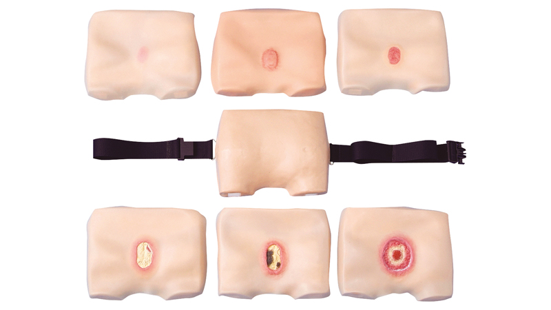
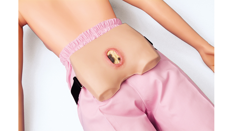
This model is a reproduction of decubitus (a bedsore) on the sacral region shown in separate stages (Stage Ⅰ to Ⅳ). It can be fitted to a human or a training model.
- Six layers of skin are used to represent Stage I to IV to enable understanding of the classification of each stage.
- The skin is mode of silicone rubber for an appearance and texture that is similar to that of the human body.
- All six skins can be layered on together and peeled off one by one to show the progression of decubitus at a glance. (Picture-card style)
- Ointment can be applied and surgical treatment can be performed.
* Do not apply ointment to the fitting holder. The surface paint may peel off.
Classification of Each Stage
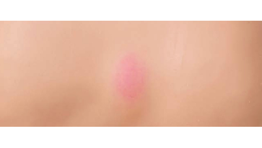
Stage Ⅰ
Circumscribed skin flare
No changes to pale skin by pressure
No injury on epidermis
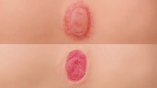
Stage Ⅱ
Partial defect of skin including epidermis and dermis
Blister and erosion are observed
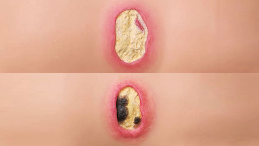
Stage Ⅲ
Defect reach to subcutaneous tissue
Sometimes pocket is formed
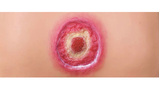
Stage Ⅳ
Deep defect down to the muscle, bone and support tissue
Pocket is formed, and sometimes surgery is required for the treatment
| Fitting holder | 1 |
|---|---|
| Stage Ⅰ Skin (flare) | 1 |
| Stage Ⅱ Skin (blister) | 1 |
| Stage Ⅱ Skin (epidermolysis) | 1 |
| Stage Ⅲ Skin (decubitus with white necrotic tissue) | 1 |
| Stage Ⅲ Skin (decubitus with black necrotic tissue) | 1 |
| Stage Ⅳ Skin (decubitus with exposed bone) | 1 |
| Baby powder | 1 |
| Size | Approx. 18(L) × 22(W) × 5(H) cm |
|---|---|
| Weight | Approx. 1.4 kg |
| Materials |
Holder: Urethane foam Skin: Silicone |

